


Can You Buy Options on Gold? Everything You Need to Know
Is it possible to purchase options on gold? Gold has been a valuable and sought-after asset for centuries. It has served as a store of value, a medium …
Read Article
An electrocardiogram (ECG) is a valuable diagnostic tool used to assess the electrical activity of the heart. It provides crucial information about the heart’s rhythm and can help identify various cardiac conditions. One important aspect of an ECG reading is the T wave, which represents repolarization of the ventricles. Normally, the T wave appears as a smooth, rounded wave.

However, in some cases, the T wave can present as notched or bifid, meaning it has a double peak or a notch in the middle. This abnormality can indicate several underlying cardiac issues and should not be disregarded. It is important for healthcare professionals to understand the significance of a notched T wave and its potential implications.
A notched T wave can be indicative of electrolyte imbalances, such as hypokalemia or hyperkalemia, which affect the heart’s electrical conduction system. It can also be an early sign of myocardial ischemia, where the heart muscle doesn’t receive enough blood flow. Additionally, a notched T wave can be associated with certain medications, like antiarrhythmic drugs or psychotropic medications.
In some cases, a notched T wave may be a benign finding and not associated with any pathological condition. It can occur in healthy individuals, especially children and young adults. However, it is essential to differentiate between a normal variant and a pathological finding, as the latter requires further investigation and management.
Read Also: Screening Forex Pairs: A Guide to Choosing the Right Currency Pairs for Trading
In conclusion, understanding the significance of a notched T wave in ECG readings is crucial for accurate interpretation and diagnosis. It can be a subtle yet important indicator of underlying cardiac issues or electrolyte imbalances. Healthcare professionals must be vigilant in recognizing this abnormality and conducting further investigations when necessary to ensure optimal patient care.
The T wave is a component of the electrocardiogram (ECG) that represents the repolarization of the ventricles of the heart. It is an important waveform that provides valuable information about the electrical activity of the heart.
A notched T wave is a variation of the normal T wave morphology where the peak of the wave is bifid or has two distinct peaks. This abnormality can be observed in certain cardiac conditions and can provide important clues for evaluating the health of the heart.
One possible cause of a notched T wave is the presence of electrolyte imbalances, specifically hypokalemia or low levels of potassium in the blood. This can disrupt the normal repolarization process and result in changes to the T wave morphology. Other causes include certain medications, such as certain antiarrhythmics or anti-ischemic drugs.
In some cases, a notched T wave may be a sign of an underlying cardiac condition. For example, it can be observed in patients with long QT syndrome, which is a disorder of the electrical system of the heart that can increase the risk of life-threatening arrhythmias. Other conditions that can cause a notched T wave include ischemia, myocardial infarction, and cardiac hypertrophy.
It is important for healthcare professionals to recognize and understand the significance of a notched T wave in ECG readings. By identifying this abnormality, appropriate diagnostic tests can be performed to further evaluate the patient’s cardiac health and determine the underlying cause of the abnormal T wave morphology.
In conclusion, a notched T wave is an abnormal variant of the T wave morphology that can indicate electrolyte imbalances or underlying cardiac conditions. Healthcare professionals should be vigilant in recognizing this abnormality and further investigating its significance in order to provide appropriate care for the patient.
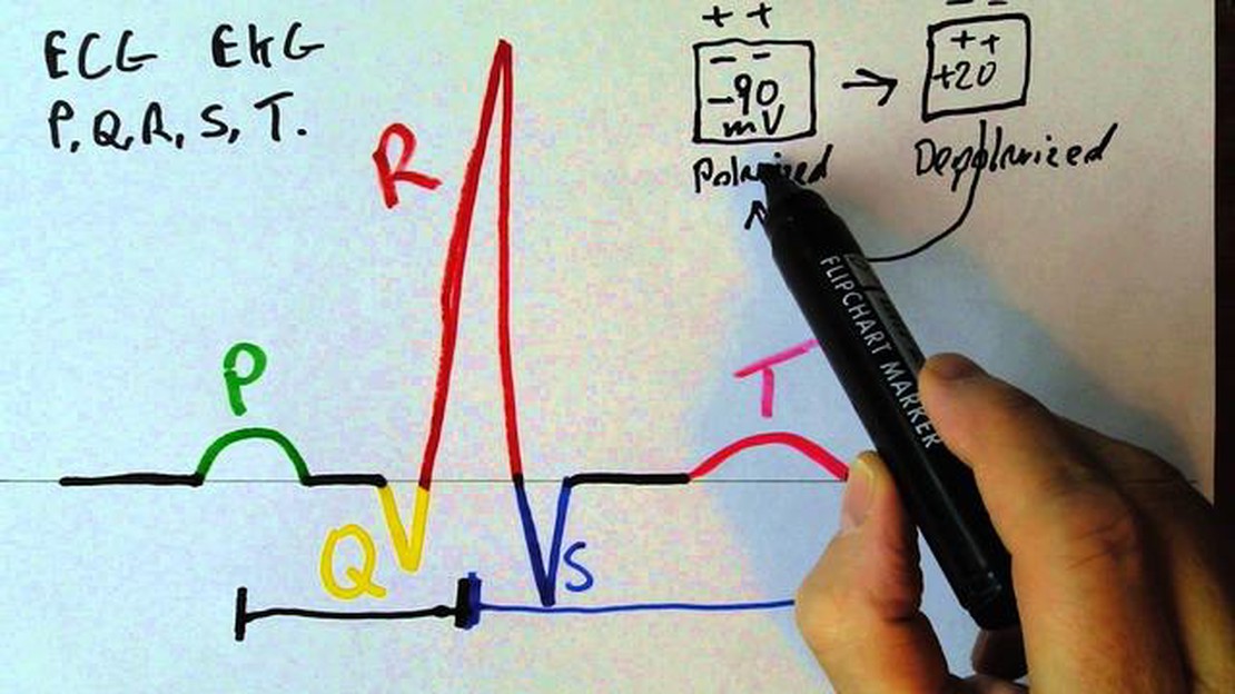
An electrocardiogram (ECG) is a diagnostic test that records the electrical activity of the heart. It is a useful tool for assessing the health of the heart and diagnosing various heart conditions. An ECG reading provides valuable information about the heart’s rhythm, rate, and overall function.
Read Also: Discover the Top Momentum Indicator for MT4 That Will Boost Your Trading Performance
The ECG reading is obtained by placing electrodes on specific points of the patient’s body, which are then connected to a machine that records the electrical impulses generated by the heart. These electrical impulses are represented as waves that are displayed on a graph.
The ECG reading consists of various waves, intervals, and segments, each of which provides specific information about the heart’s activity:
| Wave/Interval/Segment | Description |
|---|---|
| P wave | Represents the depolarization of the atria, or the contraction of the atria as they pump blood into the ventricles. |
| QRS complex | Represents the depolarization of the ventricles, or the contraction of the ventricles as they pump blood out of the heart. |
| T wave | Represents the repolarization of the ventricles, or the recovery phase of the ventricles as they prepare for the next heartbeat. |
| PR interval | Measures the time it takes for the electrical impulse to travel from the atria to the ventricles. |
| QT interval | Measures the time it takes for the ventricles to depolarize and repolarize. |
| ST segment | Represents the period when the ventricles are contractThe Importance of Notched T WavesNotched T waves are significant findings that can indicate underlying cardiovascular issues. These abnormalities can be observed in electrocardiogram (ECG) readings and may suggest a variety of conditions, including coronary artery disease, myocardial ischemia, electrolyte imbalances, and drug toxicities.The presence of a notched T wave often indicates a delay in ventricular repolarization. It suggests an altered balance in the electrical impulses within the heart, potentially leading to disturbances in cardiac function. This irregularity can be a sign of atherosclerotic plaques blocking the coronary arteries or reduced blood supply to the myocardium, both of which can significantly impact heart health.Furthermore, a notched T wave can be a manifestation of electrolyte imbalances, such as hyperkalemia or hypokalemia. Increased or decreased levels of potassium in the blood can disrupt the normal electrical conduction of the heart, resulting in T wave abnormalities. Monitoring and addressing these imbalances are crucial for preventing serious cardiac events.Drug toxicities can also cause notched T waves. Certain medications, such as antiarrhythmics and psychotropic drugs, can interfere with the normal repolarization process and lead to characteristic ECG changes. Recognizing these patterns and adjusting the medication regimen accordingly is crucial to preventing potential adverse effects on the cardiovascular system.In conclusion, the presence of notched T waves on an ECG reading should not be disregarded. These abnormalities can serve as early warning signs of cardiovascular problems, including coronary artery disease, myocardial ischemia, electrolyte imbalances, and drug toxicities. Recognizing and addressing the underlying issues can significantly improve patient outcomes and overall heart health. Regular monitoring and timely intervention are essential in managing these conditions and preventing potential complications.FAQ:What is a notched T wave in ECG readings?A notched T wave in ECG readings refers to a characteristic wave pattern seen on the electrocardiogram. It is a deviation from the normal smooth, rounded shape of the T wave, and instead shows a distinct notch or double-humped appearance.What conditions can cause a notched T wave?Several conditions can cause a notched T wave, including electrolyte imbalances, myocardial infarction, myocarditis, ventricular hypertrophy, and certain medication side effects. It is important to evaluate the clinical context and other ECG findings to determine the specific cause.What does a notched T wave indicate?A notched T wave may indicate abnormalities in ventricular repolarization. It can be a sign of an underlying cardiac condition or an electrolyte imbalance. Further investigation and evaluation are necessary to determine the significance and appropriate management.Is a notched T wave always a cause for concern?A notched T wave is not always a cause for concern. It can be a normal variant in some individuals, particularly athletes. However, it can also be indicative of an underlying cardiac abnormality or electrolyte disturbance. It is important to consult with a healthcare professional for proper evaluation and interpretation of the ECG findings.How is a notched T wave treated?The treatment for a notched T wave depends on the underlying cause. If it is related to an electrolyte imbalance, correcting the imbalance may resolve the issue. If it is associated with a cardiac condition, appropriate management strategies will be determined by a healthcare professional based on the specific diagnosis. |


Is it possible to purchase options on gold? Gold has been a valuable and sought-after asset for centuries. It has served as a store of value, a medium …
Read Article
Is it Possible to Day Trade 3 Times a Week? Day trading is a popular form of trading where traders open and close positions within the same day to …
Read Article
Calculating Your Take-Home Pay: How Much is $90000 after Taxes in California? Living in California can be expensive, but it also has its benefits. As …
Read Article
Python for Technical Analysis: A Comprehensive Guide Technical analysis is a method used by traders and investors to analyze and predict future price …
Read Article
Understanding Stock Put Options: What You Need to Know A stock put option is a financial contract that gives the holder the right, but not the …
Read Article
Taxation on Binary Trading: Understanding the Rules and Regulations Binary trading, also known as digital options or fixed-return options, has gained …
Read Article


