


Is IQ Option Regulated? Exploring the Regulation of IQ Option
Is IQ Option regulated? When it comes to choosing a trading platform, it’s important to consider whether it is regulated or not. Regulation ensures …
Read Article
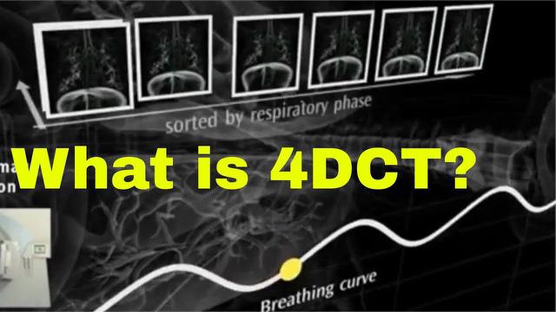
When it comes to medical imaging, CT (Computed Tomography) scans are a standard diagnostic tool. They provide detailed cross-sectional images of the body, allowing doctors to detect and diagnose various conditions. However, traditional CT scans capture images of the body in a static state, which can limit their effectiveness in capturing dynamic processes, such as breathing or motion of organs.
This is where 4D CT simulation comes into play. 4D CT simulation is an advanced imaging technique that combines the power of CT scanning with the ability to capture and analyze motion. It uses a series of images taken over time to create a 4D model, allowing doctors to visualize anatomical structures in motion and study how they change over time.
One of the major applications of 4D CT simulation is in radiation therapy planning. It allows radiation oncologists to precisely target tumors while minimizing damage to healthy tissues. By taking into account the motion of organs during treatment, 4D CT simulation helps ensure accurate delivery of radiation and improves treatment outcomes.
In addition to radiation therapy planning, 4D CT simulation has other important medical applications. It can be used to study respiratory function and lung diseases, analyze cardiac function, and assess the movement of other organs such as the liver or gastrointestinal tract. The ability to visualize and analyze motion in real-time provides valuable insights for diagnosis, treatment planning, and monitoring of various medical conditions.
In conclusion, 4D CT simulation is revolutionizing medical imaging by capturing and analyzing motion. It allows doctors to study anatomical structures in real-time, enabling more accurate diagnoses and treatment planning. From radiation therapy to respiratory function, 4D CT simulation has numerous applications in the medical field, helping improve patient care and outcomes.
4D CT simulation is a medical imaging technique that allows healthcare professionals to generate images of the body in real-time and track the movement of internal structures. It is commonly used in radiation therapy planning for cancer treatment.

4D CT simulation works by combining computed tomography (CT) scanning with synchronized patient respiratory data. During the scan, the patient is asked to breathe normally or follow a specific breathing pattern, while the CT scanner takes a series of images.
Read Also: Scalping Forex with Spread: A Guide to Effective Trading Strategies
Here’s how the process works:
Overall, 4D CT simulation provides valuable information about how organs and tumors move during breathing, which helps in delivering precise radiation therapy treatment. It allows the radiation therapy team to account for motion and deliver radiation to the tumor while minimizing damage to healthy tissues.
4D CT simulation, also known as four-dimensional computed tomography simulation, is a powerful tool used in medical imaging that offers several benefits for both patients and healthcare professionals. This advanced imaging technique combines the capabilities of traditional CT scanning with the ability to visualize moving organs and tissues in real-time.
One of the major advantages of 4D CT simulation is its ability to accurately capture and analyze dynamic movements within the body. By acquiring a series of images at different time points, doctors can observe how structures such as lungs, heart, and diaphragm move and interact during respiration. This level of detail allows for more precise treatment planning and delivery, particularly in radiation therapy.
Another benefit of 4D CT simulation is its ability to reduce uncertainties in treatment planning. Because it provides a comprehensive understanding of the patient’s anatomy and organ motion, healthcare professionals can more accurately define the treatment target and spare healthy surrounding tissues from unnecessary radiation exposure. This leads to better treatment outcomes and reduced side effects for patients.
In addition, 4D CT simulation offers valuable insights into vascular structures, making it an essential tool in diagnosing and treating cardiovascular diseases. It allows physicians to study blood flow and identify potential blockages or abnormalities in real-time. This information is vital for assessing cardiovascular conditions, planning interventions, and guiding surgery.
Furthermore, 4D CT simulation is a non-invasive imaging technique that minimizes patient discomfort and risk. Unlike invasive procedures, such as angiography, 4D CT simulation does not require the insertion of catheters or contrast agents, reducing the potential for complications. Patients can undergo the procedure without anesthesia and experience minimal downtime, contributing to a more efficient and patient-friendly healthcare experience.
Overall, 4D CT simulation offers numerous benefits in medical imaging. Its ability to capture dynamic movements, reduce uncertainties in treatment planning, and provide valuable insights into vascular structures makes it an indispensable tool in various medical fields. The continued advancements in 4D CT technology will likely lead to further improvements in healthcare outcomes and patient care.
Read Also: Understanding Trading Alerts: What They Are and How They Work
4D CT simulation is a medical imaging technique that combines CT scanning with motion tracking to capture dynamic images of organs in motion. It allows doctors to better understand and plan treatments for conditions that involve movement, such as lung cancer.

During a 4D CT simulation, the patient lies on a CT table while a series of CT scans is taken at different time points. These scans are synchronized with the patient’s breathing or other movements to capture the organ in motion. The images from each time point are then combined to create a 4D image sequence, which can be played back to visualize the motion.
4D CT simulation offers several advantages over traditional CT scanning. It provides a more accurate representation of motion compared to static images, allowing doctors to better target treatments. It also reduces the need for invasive procedures, as it can provide valuable information about tumor motion without the need for additional tests.
4D CT simulation is commonly used in radiation therapy planning for conditions such as lung cancer, where tumor motion can be a significant factor in treatment effectiveness. It is also used in other areas of medicine, such as cardiology, to evaluate heart function and blood flow during different phases of the cardiac cycle.
Like any medical procedure, there are some risks associated with 4D CT simulation. However, the risks are generally minimal. The radiation exposure from a CT scan is slightly higher than that of a conventional X-ray, but it is still considered safe. There may also be some discomfort or anxiety related to lying still for an extended period of time during the procedure.

Is IQ Option regulated? When it comes to choosing a trading platform, it’s important to consider whether it is regulated or not. Regulation ensures …
Read Article
FXOpen: Is the Platform Open for US Clients? Yes, US clients can trade with FXOpen! FXOpen is a global forex and CFD broker that offers trading …
Read Article
Customizing your Moving Average in Tradingview TradingView is a powerful platform for traders to analyze and visualize financial markets. One of the …
Read Article
Are non qualified stock options reported on W2? If you have received non-qualified stock options (NSOs) from your employer as part of your …
Read Article
Predictions for the Currency Exchange Rate in 2023 As we approach the year 2023, financial analysts and economists have started making predictions …
Read Article
Best Places to Exchange Currency in Uruguay When traveling to Uruguay, it’s important to know where you can exchange your currency for the best rates. …
Read Article


